


Pain in the hip jointIs specific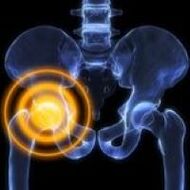 unpleasant and unbearable sensations due to pathology of the upper femur, acetabulum, adjacent soft tissue structures. In terms of intensity, they vary from weak to unbearable, in nature they can be dull, sharp, pressing, sore, broken, drilling, and others. Often they depend on load, time and other factors. The cause of pain was determined using X-rays, CT, MRI, ultrasound, arthroscopy, and other studies. Pain relievers and limb rest are recommended until a diagnosis is made.
Causes of pain in the hip joint
Soft tissue injuries
The most common cause of pain trauma is the spread of the hip joint. It occurs when falling on the side or a direct impact, manifests itself in moderate acute pain, which quickly becomes dull, gradually diminishes and disappears in a few days, in severe cases-weeks. Support is taken care of, his movement is slightly limited. Edema is detected locally, bruising may occur.
Injuries to the ligaments of the hip joint are rare, usually as a result of road accidents and sports injuries, accompanied by severe pain, sometimes - a cracking sensation (such as from torn tissue). The pain subsides slightly, then often increases again due to edema. Swelling from the joints extends to the groin area, thighs.
The degree of traumatic dysfunction of the ligament apparatus depends on the severity of the injury (stretch, tear, rupture), ranging from slight limitation to an inability to support the foot. The pain increases with deviation of the trunk, movement in the opposite direction of the damaged ligament.
Bone and joint injuries
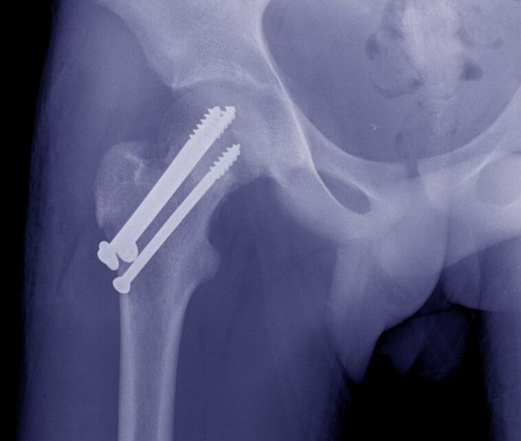
Hip fractures usually occur in the elderly as a result of domestic or street trauma. A special feature, especially when there is osteoporosis, is the absence of intense pain syndrome, mild edema. At rest, the pain is deep, dull, moderate or insignificant, with the movement of the painful sensation increasing sharply. Support is sometimes maintained. A common symptom is an inability to lift a straightened leg from a prone position (symptoms of a stuck heel).
Transtrochanteric fractures are more frequently diagnosed in middle- and young people and develop as a result of high -energy trauma. Unlike cervical fractures, they are accompanied by deep, diffuse pain that is unbearable. Then the pain subsided, but remained strong, unbearable. Swollen joints, bruising may occur. Movement is very limited. Support is not possible.
Isolated fractures of the larger trochanter are rare; it is found in children and young people; it is formed by a fall, direct impact, or sharp muscle contraction. The pain is acute, very intense, localized mainly on the outer surface of the joint. Because of the increased pain, the patient avoids active movement.
Hip dislocation occurs when falling from a height, industrial and road traffic injuries, manifested in unbearable sharp pain that almost does not subside until reduction. Defective joints, legs shortened, bent at the knee joint, turned outwards, more rarely inwards (depending on the type of dislocation). Support and movement are not possible, when trying to move, spring resistance is determined.
Acetabular fractures develop separately or in combination with hip dislocation. Characterized by acute explosive pain in the depths of the hip joint. After that, the pain subsided, but remained strong, preventing any movement. Legs shortened, rotated outwards. Support is not possible.
Degenerative process
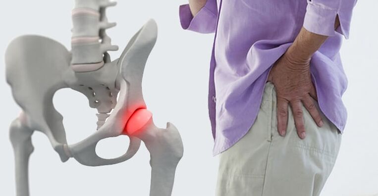
With coxarthrosis in the early stages, periodic pain, dull, not localized, appearing at the end of the day or after a significant load, sometimes radiating to the hip, knee joints. Light and fast stiffness can be done at the beginning of the movement. After that, the intensity of pain increases, painful sensations are observed not only during movement, but also during rest. After struggling, the patient began to drown. Movement is quite limited.
In severe coxarthrosis, the pain is felt deep, diffuse, persistent, aching, twisting. Disturb both day and night. Resistance to stress is reduced; when walking, the patient leans on a cane. Movement is significantly limited, the affected leg is shortened, which leads to increased load on the joints, increased pain when walking and standing.
Chondromatosis of the hip joint in its course resembles subacute arthritis. The pain is moderate, diffuse, temporary, combined with cracking, limitation of movement. When the intra-articular body is violated, blockage occurs, characterized by intense sharp pain, impossibility or significant limitation of movement. After cessation of articular mouse violations, the listed symptoms disappeared.
Trochanteritis usually forms with arthrosis of the hip joint, accompanied by inflammatory-degenerative lesions on the tendons of the gluteal muscles at the point of its attachment to the larger trochanter, indicated by pain in the lesion area at supine position on the affected part. There is an increase in pain when trying to abduct the hip with endurance.
Bone nutrition disorders
Perthes disease develops in children and adolescents, characterized by partial necrosis of the femoral head, which is initially accompanied by mild to moderate pain, sometimes radiating to the knees and hips. After a few months, the pain gets stronger, persistent, sharp, and exhausting. The joint swells, its movement is limited, and lameness occurs. Then the pain subsides, the degree of recovery of joint function varies.
Aseptic necrosis of the downstream femoral head resembles Perthes disease, but is detected in adults, walking poorly, in half of the cases bilateral. At first, the pain is periodic, interesting. Then the pain syndrome worsens, appearing at night. At the peak of clinical manifestations, the pain is so intense that the person completely loses the ability to lean on his feet. Then the pain gradually subsides. Restriction of movement progress for about 2 years, the result is arthrosis of the hip joint, contractions, and shortening of the limbs.
Solitary bone cysts form on the metaphysis of the proximal thigh in boys aged 10–15 years, accompanied by mild intermittent pain in the hip joint. Edema is usually absent, with prolonged contractions often occurring, especially in young children. Due to mild symptoms, the cause of treatment is a pathological fracture or an increasing limitation of movement.
Arthritis
Aseptic arthritis is indicated by wave -like pain in the joints, which increases in the morning. Pain severity varies from insignificant to acute, intense, persistent, significantly limiting physical activity. Stiffness, swelling, redness, and an increase in local temperature were observed. Palpation is painful.
In rheumatoid arthritis, the hip joint is rarely involved, the lesions are symmetrical. Periodic pain first appears against the background of changing seasons (autumn, spring), with sudden changes in weather conditions, during hormonal changes after childbirth or during menopause. The pain is moderate or weak, diffuse, pulling or aching, increasing sharply on palpation. It is combined with recurrent synovitis, edema, hyperemia, hyperthermia, increased range of motion.
Infectious arthritis develops with the spread of hematogenous or lymphogenic infections, more rarely - with the penetration of pathogens into the joints from nearby tissues. Usually the acute onset with rapidly increasing painThe pain feels intense, wrinkled, tearing, ruptured, disturbed at rest, exacerbated by movement, as the limb assumes a forced position. The patient experienced fever, chills, sweating, severe weakness, edema, redness in the joints, and an increase in local temperature.
If there is no timely treatment, bacterial infectious arthritis can turn into panarthritis - a purulent inflammation of all the tissues of the hip joint. It is characterized by a severe course with very acute throbbing pain, hectic fever, severe weakness, pre-syncope, hyperemia and marked hyperthermia.
Other inflammatory diseases
Upper thigh osteomyelitis can be hematogenous, post -traumatic, or postoperative. Hematogenic osteomyelitis is manifested by rupture, cramping, tearing, or very acute pain, which causes the patient to avoid limb movement in the slightest. There is marked hyperthermia, severe intoxication.
Post -traumatic and postoperative osteomyelitis occur with similar, but less pronounced, symptoms. Typically, a more gradual onset against the background of open fractures or postoperative wounds, the appearance of purulent discharge. Pain in the hip joint increases in 1-2 weeks in parallel with the development of signs of local inflammation.
Synovitis develops against the background of injuries, other diseases of the hip joint, the more rarely it becomes a manifestation of allergies. In acute synovitis, the pain is usually mild, dull, ruptured, increasing gradually due to an increase in the amount of intra-articular fluid. The joints are swollen, palpation is slightly painful, the symptoms of fluctuations are determined. Chronic synovitis is asymptomatic, accompanied by mild pain.
With intermittent hydrarthrosis, the pain is also insignificant, accompanied by discomfort, limited movement, and disappears within 3-5 days after reabsorption of reverse effusion. They are renewed periodically, individually for each patient, due to the repeated accumulation of fluid in the joints.
Specific infections
Tuberculosis of the hip joint is a common form of osteoarticular tuberculosis, which manifests itself with general weakness, fatigue, subfebrile conditions. Then there is an excruciating pain or soreness in the muscles, while a painful sensation in the joints when walking. The patient begins to stretch the limbs. As the pain increases, they become moderate, diffuse, radiating to the knee, coupled with swelling, redness, synovitis. Protective contractors thrive.
Joint pain, including hip pain, may appear with brucellosis. In acute and subacute forms, painful sensations pull, twisting, combined with periodic fever, lymphadenopathy, skin rash. In the chronic course, the pain syndrome resembles that in aseptic arthritis, a deformity over time forms.
Congenital anomalies
The manifestation of hip dysplasia is determined by the degree of imbalance of the femoral head and acetabulum. With complete congenital dislocation, pain appears as soon as the child begins to walk, accompanied by lameness. With moderate subluxation, a painful sensation occurs at the age of 5-6 years, directly related to the load on the legs.
With mild subluxation, asymptomatic pathology for a long time, pain syndrome manifests itself with the development of dysplastic coxarthrosis at the age of 25-30 years. The peculiarities of such arthrosis are the rapid intensification of pain, the onset of pain at rest and at night, and the progressive limitation of movement. All forms of dysplasia are accompanied by asymmetry of skin folds, "click" symptoms and limited movement. If dislocated, limb shortening is observed.
Neoplasm
For benign neoplasms, a typical asymptomatic course. The pain is mild, intermittent, and often does not increase for years. Tumor growth is accompanied by an increase in slow pain syndrome, recurrent synovitis. In the area of the hip joint, osteoma, osteoid osteoid, osteoblastoma, chondromas are more frequently detected.
Malignant neoplasias (osteosarcomas, chondrosarcomas) are characterized by the rapid development of pain syndromes and other pathological manifestations. At first, the pain is mild, short-lived, without specific localization, sometimes worse at night. After that, they become sharp, permanent, cut, surround, spread throughout the joint. The affected area is swollen, deformed. Weight loss, weakness, subfebrile conditions were observed. With advanced neoplasms, the excruciating and unbearable pain is relieved only with narcotic drugs.
Another reason
Pain in the hip joint sometimes appears with lumbosacral plexitis and sciatic nerve neuropathy, however, they usually occupy an insignificant position in the clinical picture of the disease, fading into the background compared to strong pain in the back and thighs, limb weaknessand sensitivity disorders.
This localization pain syndrome is often detected in osteochondrosis and disc herniation. Pain can be detected with spondylitis, deformed spondyloarthrosis and curvature of the spine. The pain is dull, periodic, aching, often increasing during the period of the underlying disease. The cause of their appearance can be a constant joint strain or the development of coxarthrosis.
Sometimes joint pain is triggered by mental illness or a depressive disorder. Diabetes mellitus is often accompanied by enthesopathy, capsulitis, and other lesions of the periarticular soft tissues. Possibility of arthropathy medication while taking certain medications.
Diagnostics
In the event of an injury, diagnostic measures are performed by a traumatologist. Degenerative and inflammatory diseases are managed by orthopedics and rheumatologists. If the process is purulent, the participation of a surgeon is required. Examination includes collection of complaints, review of medical history, physical examination, additional research. Taking into account the uniqueness of the pathological process, the following methods can be used:
- Radiography.This is a basic technique for most joint diseases. Detects fractures, dislocations, changes in the contour of the acetabulum and femoral head, marginal and intraosseous deformities, bone growth, narrowing of the joint space.
- Ultrasound.Most informative when studying soft tissues. Reveals signs of inflammatory and degenerative processes, areas of calcification. Used to diagnose effusion, articular rats.
- MRI and CT.Clarification techniques are used if there are unclear data from baseline studies, to clarify the nature, prevalence and location of pathological foci. Can be run with contrast.
- Joint puncture.Has a diagnostic or therapeutic and diagnostic character. Allows you to eliminate effusions, study the composition of intra-articular fluid, determine the causative agent of infection using laboratory tests.
- Arthroscopy.During the visual examination of the joint, the doctor assesses the condition of the bone and soft tissue structures, if necessary, takes biopsy samples for subsequent histological examination, and performs therapeutic measures.
- Lab test.They are prescribed to determine the signs of inflammation and markers of rheumatological diseases, to assess the general condition of the body, the activity of various organs in severe infectious or systemic pathology.
Treatment
Please before diagnosis
In severe injuries, it is necessary to correct the joint by applying fragments from the foot to the armpit. If the trauma injury is minor, it is enough to give a rest to the leg. Cold is applied to the affected area. For severe pain, analgesics are given. You can not do active movements with the limbs, loading the legs. It is strictly forbidden to attempt to remove dislocations or bone transplants.
Tactics for non -traumatic illness are determined by symptoms. With minor manifestations, it is allowed to keep the whole limb, the use of local drugs with analgesic and anti-inflammatory effect. If fever, weakness, severe pain, edema and rapid hyperemia, it is recommended to immediately seek specialized help.
Conservative therapy
Dislocation is an indication for immediate reduction. If a fracture, skeletal traction is usually used, then the patient is operated on or the limb is fitted with a cast plaster after signs of callus appear. In elderly patients with hip fractures, immobilization with derotation boots is allowed, which prevents rotational movement of the joints.
The rest of the patient is advised to relieve the hip joint. According to the instructions, it is recommended to use orthoses or additional tools (sticks, canes). Determine the sequence, physiotherapy exercises, physiotherapy procedures:
- laser therapy;
- magnetotherapy;
- UHF;
- ultrasound;
- electrophoresis with drugs;
- UHT.
Possibility of using NSAIDs, chondroprotectors, antibacterial drugs. Local agents are widely used. According to the indications, joint puncture, intra- and periarticular obstruction with hormones, intra-articular injection of chondroprotectors, synovial fluid replacement were performed.
Surgery
Surgery on the hip joint is performed with open access and with the help of arthroscopic equipment. Taking into account the type of pathology, the following can be done:
- Traumatic injuries:open reduction of hip dislocation, acetabulum remodeling, neck osteosynthesis, trochanteric fractures.
- Degenerative process:arthrotomy, arthroscopy, removal of independent intra-articular body.
- Growth:removal of neoplasia, bone resection, disarticulated hip joint, Io-abdominal amputation, Io-abdominal resection.
In the event of contractions, ankylosis, scarring of periarticular tissue, rehabilitation, arthroplasty, and arthrodesis are performed. Endoprosthetics are an effective method of restoring limb function in diseases of various origins, accompanied by limitation of movement or joint damage.